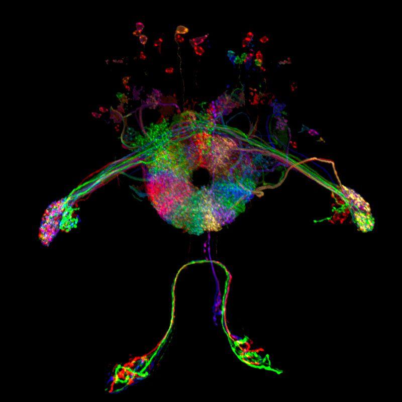
The doughnut–shaped ring at the centre of the image below shows the set of “compass neurons”
recently detected in a fruit fly’s brain. Each coloured segment in the ring represents a separate neuron;
together, they make up the “neural compass” that allows a fly to quickly establish a sense of direction in
a new environment. The compass neurons are connected to each other, and only allow one neuron to
be active at a given time. They are also linked to visual neurons that are bringing information from the
fly’s eyes. Together, the visual cues and the compass neurons provide the fly with the information
necessary to orient itself, and choose which direction to fly.

Research Article Title
Sensorimotor experience remaps visual input to a heading-direction
network
Y. Fisher et al , Nature (Nov. 20, 2019) and Campbell and Giocomo, Nature News and Views
(Nov.20, 2019)
As everyone knows, a good sense of direction is needed to successfully navigate the world. In
mammals, this ‘sense’ involves neurons called head-direction cells. Each such cell becomes
most active when the animal faces a particular direction relative to landmarks in its
environment. Together, the cells’ activity indicates which direction the animal is facing at any
given moment. In 2015, it emerged that fruit flies (which are much easier than mammals to
study experimentally) have strikingly similar cells, called heading neurons (1) . Writing in Nature,
Fisher et al. (2) and Kim et al. (3) now build on this discovery to tackle a decades-old problem: how
does this type of neuron respond to the locations of landmarks in a manner that is stable
enough to be reliable, but flexible enough to allow adaptation to new environments?
To give an example of the problem, imagine emerging from a subway station onto a crowded
street. If you are a regular visitor, a glance around is all you need to be on your way. However, if
you have never been to this station before, you might need a moment to orient yourself. You
take note of surrounding street signs, shops and monuments. Before long, you have your
bearings and can set off in the right direction.
This example highlights two challenges for the brain’s directional system. First, it must stably
indicate direction in familiar environments: returning to the same station should call the same
orientation to mind. Second, it must have the flexibility to learn new configurations of
landmarks, even when similar landmarks have been seen before — the particular configuration
of street signs at the new station must be learnt, even though you may have seen similar street
signs in other places.
The neural mechanisms that underlie these abilities in flies are a beautiful example of form
following function. The insects’ heading neurons (also known as… compass neurons) are
arranged in a ring (see image above) that corresponds to the 360° of possible directions in
which the fly can face, sometimes called heading angles. Because of inhibition between
neurons, only one heading angle can be indicated at one time, providing the fly with an
unambiguous [directional] signal. Of note, rather than always aligning their activity to a cardinal
direction such as north, heading neurons realign their activity arbitrarily when the fly enters a
new environment. The heading neurons receive input from visual ring neurons, which are
activated by visual cues at particular orientations relative to the fly, and from internal cues
about self-motion.
 Orienteering BC